
Introduction
Dental imaging plays a crucial role in diagnosing and treating various dental conditions. Over the years, there have been significant advancements in dental imaging technology, leading to safer and more accurate diagnostics. These advancements have revolutionized the field of dentistry, allowing dentists to provide better care and treatment options for their patients. In this article, we will explore some of the latest advances in dental imaging and how they have improved diagnostics.
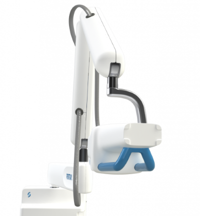
Cone Beam Computed Tomography (CBCT)
Cone Beam Computed Tomography, or CBCT, is a revolutionary imaging technique that provides three-dimensional images of the oral and maxillofacial region. Unlike traditional dental X-rays, CBCT offers a more comprehensive view, allowing dentists to assess bone density, detect abnormalities, and plan complex dental procedures with precision. The use of CBCT has significantly improved the accuracy of dental diagnoses and treatment planning.
Digital Radiography
Digital radiography has replaced traditional film-based X-rays in many dental practices. This technology uses electronic sensors to capture and store dental images digitally. Digital radiography offers several advantages, including reduced radiation exposure, instant image availability, and the ability to enhance and manipulate images for better diagnostics. Dentists can now zoom in, adjust contrast, and highlight specific areas of interest, leading to more accurate diagnoses.
Intraoral Cameras
Intraoral cameras are small, handheld devices that allow dentists to capture high-resolution images of the inside of a patient’s mouth. These cameras provide a detailed view of teeth, gums, and other oral structures, helping dentists identify dental issues that may not be visible to the naked eye. Intraoral cameras also enable dentists to educate patients about their oral health by showing them real-time images, fostering better communication and understanding.
Digital Smile Design (DSD)
Digital Smile Design is a cutting-edge technology that combines dental imaging with computer software to create a digital representation of a patient’s smile. Dentists can use DSD to design and simulate various dental procedures, such as veneers, crowns, and orthodontic treatments, before actually performing them. This technology allows patients to visualize.
Summary
The latest advances in dental imaging have brought about remarkable improvements in diagnostic capabilities. With the introduction of digital radiography, traditional film-based X-rays have been replaced by digital sensors, reducing radiation exposure for patients. This technology also allows for enhanced image quality, making it easier for dentists to detect and diagnose dental issues.
Furthermore, cone beam computed tomography (CBCT) has emerged as a game-changer in dental imaging. CBCT provides three-dimensional images of the teeth, jawbone, and surrounding structures, enabling dentists to accurately plan and execute complex procedures such as dental implant placement and orthodontic treatments.
Another significant advancement is the development of intraoral scanners, which eliminate the need for messy and uncomfortable traditional dental impressions. These scanners capture highly detailed digital impressions of the patient’s teeth and gums, allowing for precise treatment planning and the creation of custom restorations.
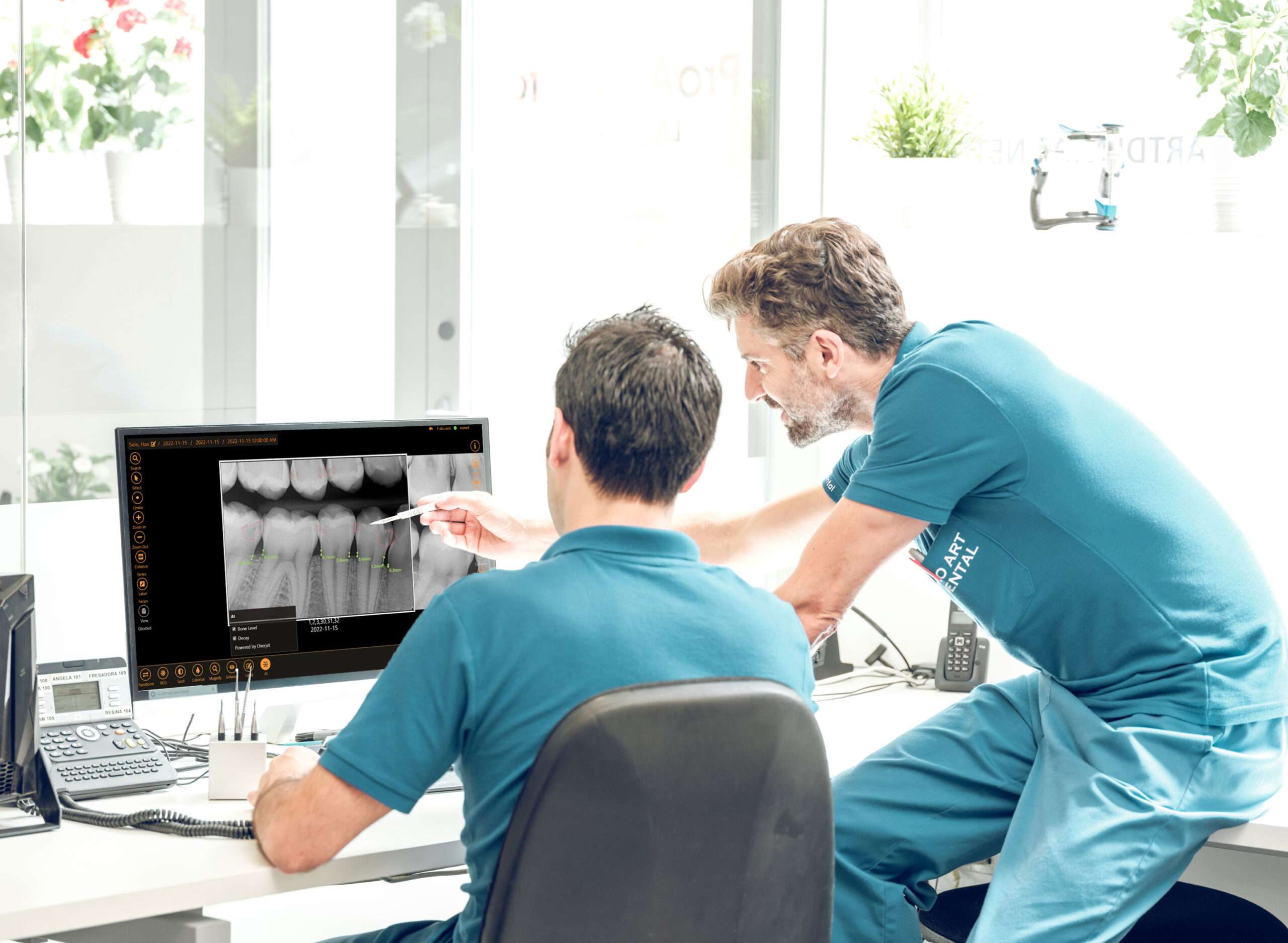
Additionally, the integration of artificial intelligence (AI) in dental imaging software has further improved diagnostics. AI algorithms can analyze dental images and assist dentists in identifying abnormalities or potential issues that may have been overlooked. This technology helps in early detection and timely intervention, leading to better treatment outcomes.
In conclusion, the latest advances in dental imaging have revolutionized the field of dentistry, providing safer and more accurate diagnostics. With digital radiography, CBCT, intraoral scanners, and AI-powered software, dentists can now detect and treat dental problems with greater precision and efficiency. These advancements ultimately contribute to improved pat ient care and overall oral health.
- Q: What are the latest advances in dental imaging?
- A: The latest advances in dental imaging include technologies such as cone beam computed tomography (CBCT), digital radiography, and intraoral scanners.
- Q: How do these advances make diagnostics safer?
- A: These advances make diagnostics safer by reducing the amount of radiation exposure compared to traditional X-rays, while still providing high-quality images for accurate diagnosis.
- Q: How do these advances improve diagnostic accuracy?
- A: These advances improve diagnostic accuracy by providing detailed 3D images, allowing dentists to visualize dental structures from different angles and detect potential issues that may not be visible with 2D imaging.
- Q: What is cone beam computed tomography (CBCT)?
- A: CBCT is a technology that uses a cone-shaped X-ray beam to capture detailed 3D images of the teeth, jawbone, and surrounding structures. It provides valuable information for dental implant planning, orthodontic treatment, and detecting abnormalities.
- Q: What is digital radiography?
- A: Digital radiography is a technique that replaces traditional X-ray films with digital sensors. It allows for instant image acquisition, manipulation, and sharing, reducing the need for chemical processing and improving efficiency.
- Q: What are intraoral scanners?
- A: Intraoral scanners are devices used to capture digital impressions of a patient’s teeth and oral tissues. They eliminate the need for messy traditional impressions, provide more comfort to patients, and enable precise measurements for restorations and orthodontic treatments.

Welcome to my website! My name is Dr. Andrew Prinsep, and I am thrilled to share my passion for aesthetic dentistry, Invisalign and braces, dental checkups, and oral cancer screenings with you.
As a professional dental assistant, I have dedicated my career to helping patients achieve their dream smiles and maintain optimal oral health.